Female Upper Thigh Anatomy - Female Muscle Diagram And Definitions Jacki S Blog : Muscles of the anterior thigh.. Lunges are a terrific exercise for strengthening the gluteal muscles, and also the upper thighs. There may be variations in treatment that. The final chapter presents anatomical charts of anatomical sections of the upper limb: The information contained in anatomy atlases is not a substitute for the medical care and advice of your physician. Collection by renaud galand • last updated 12 weeks ago.
Collection by renaud galand • last updated 12 weeks ago. Appendicular muscles of the pelvic girdle and lower limbs. Muscles of the leg and foot. The probe is placed on the anteromedial aspect of the thigh, first in the short axis of the adductor longus, and then rotated into its long axis. Free access interactive and dynamic anatomical atlas.
Sartorius Muscle High Resolution Stock Photography And Images Alamy from c8.alamy.com It is part of the lower limb. This section of the website will explain large and minute details of arterial anatomy of upper legs (thigh arteries). Origin:upper part of intertrochanteric line. This webpage presents the anatomical structures found on thigh mri. Stiff legged deadlifts work the muscles in the back of the legs. Lymphovenous anastomosis (lva) requires a precise knowledge of the anatomy of the superficial lymphatic collectors in relation to the superficial methods: To practice tricky questions and answers on all areas of human anatomy, here is complete set of 1000+ multiple choice questions and answers. 2, vastus medialis & intermedius muscles.
Anatomy lectures , muscles of anterior compartment of thigh.
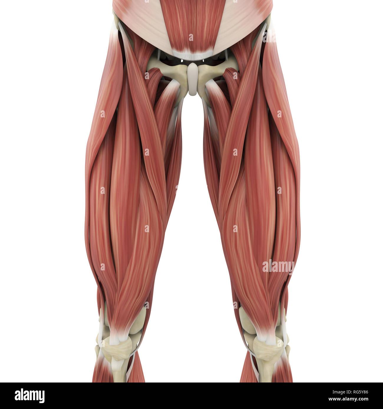
Deviantart is the world's largest online social community for artists and art enthusiasts. Quadriceps tendon into patella, then via ligamentum patellae into tubercle of tiba. This can effectively educate everyone on the female human body. It is present in upper thigh that helps blood supply to neck and head of the femur. Muscles in the medial compartment of the thigh. Move on to the muscles and bones of the thigh. The single bone in the thigh is called the femur. Appendicular muscles of the pelvic girdle and lower limbs. Our objective was to describe the muscular and neurovascular anatomy of the medial thigh compartment. These images are a random sampling from a bing search on the term thigh anatomy. click on the image (or right click) to open the source website in a new browser window. Dissections were performed in unembalmed female cadavers. The information contained in anatomy atlases is not a substitute for the medical care and advice of your physician. Upper thigh anatomy (page 1).
Muscles in the medial compartment of the thigh. Free access interactive and dynamic anatomical atlas. Anatomy lectures , muscles of anterior compartment of thigh. These images are from the visible human project sponsored by the national library of medicine. Muscles of the anterior thigh.
Top 8 Exercises To Build The Body Of A Greek God Leg Muscles Anatomy Muscle Anatomy Body Anatomy from i.pinimg.com Muscles of the leg and foot. Quadriceps tendon into patella, then via ligamentum patellae into tubercle of tiba. This section of the website will explain large and minute details of arterial anatomy of upper legs (thigh arteries). Collection by renaud galand • last updated 12 weeks ago. Appendicular muscles of the pelvic girdle and lower limbs. Anatomy lectures , muscles of anterior compartment of thigh. Move on to the muscles and bones of the thigh. Our objective was to describe the muscular and neurovascular anatomy of the medial thigh compartment.
Relationships of medial thigh structures were evaluated relative to the midpubic arch and obturator nerve.
Our objective was to describe the muscular and neurovascular anatomy of the medial thigh compartment. Adding extra weight with dumbbells while lunging can help enhance your results by toning these muscles more quickly. The single bone in the thigh region is called the femur. The information contained in anatomy atlases is not a substitute for the medical care and advice of your physician. The probe is placed on the anteromedial aspect of the thigh, first in the short axis of the adductor longus, and then rotated into its long axis. • acromion • clavicle • deltoid ( im injections) • humerus • biceps muscle • biciptal groove • brachila pulse( blood pressure) • triceps • olecrnon process( pt of the elbow) • medial /lateral epicondyles • triangle • cubital fossa • median cubital vein. Deviantart is the world's largest online social community for artists and art enthusiasts. Origin:upper part of intertrochanteric line. Anatomically, it is part of the lower limb. Vascular anatomy of the upper arm. See more ideas about female bodies, anatomy, female anatomy. Lymphovenous anastomosis (lva) requires a precise knowledge of the anatomy of the superficial lymphatic collectors in relation to the superficial methods: These images are arranged in radiographic view, as though you were looking up from the patient's feet toward the head.
Want to learn more about it? The thigh bears much of the load of the body's weight when a person is upright. Free access interactive and dynamic anatomical atlas. Muscles in the medial compartment of the thigh. Quadriceps tendon into patella, then via ligamentum patellae into tubercle of tiba.
44 Muscles And Anatomy Ideas Anatomy Muscle Anatomy Human Anatomy from i.pinimg.com Deviantart is the world's largest online social community for artists and art enthusiasts. It contains many muscles and nerves but only has one bone, the femur, which is the longest and strongest bone in the human body. Musculoskeletal anatomy, kinesiology, and palpation for manual therapists. Upper thigh anatomy (page 1). Our objective was to describe the muscular and neurovascular anatomy of the medial thigh compartment. Relationships of medial thigh structures were evaluated relative to the midpubic arch and obturator nerve. Thus, the right side of the image is the patient's left. See more ideas about female bodies, anatomy, female anatomy.
To practice tricky questions and answers on all areas of human anatomy, here is complete set of 1000+ multiple choice questions and answers.
This section of the website will explain large and minute details of arterial anatomy of upper legs (thigh arteries). The thigh is the area between the hip and the knee joint. Lymphovenous anastomosis (lva) requires a precise knowledge of the anatomy of the superficial lymphatic collectors in relation to the superficial methods: Free access interactive and dynamic anatomical atlas. The thigh bears much of the load of the body's weight when a person is upright. Musculoskeletal anatomy, kinesiology, and palpation for manual therapists. Muscles of the leg and foot. Anatomy atlases, the anatomy atlases logo, and a digital library of anatomy information are all trademarks of michael p. Anatomy lectures , muscles of anterior compartment of thigh. This webpage presents the anatomical structures found on thigh mri. Collection by renaud galand • last updated 12 weeks ago. Relationships of medial thigh structures were evaluated relative to the midpubic arch and obturator nerve. Origin:upper part of intertrochanteric line.
The single bone in the thigh is called the femur upper thigh anatomy. Origin:upper part of intertrochanteric line.


0 Comments